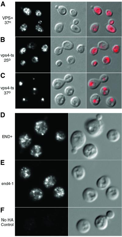
Fig. 1. Mislocalization of Arn1p in a vps4-ts strain and localization to the endosome in END+ and end4-1 strains. (A–C) Strains SEY6210 (VPS+) and MBY3 (vps4-ts) were transformed with pArn1-HA and grown in iron-poor medium at 25°C. Aliquots of culture were shifted to 37°C (A and C) for 1 h or grown at 25°C (B) prior to fixation and preparation for indirect immunofluorescence. Mouse monoclonal HA.11 was the primary antibody and Cy3-conjugated donkey anti-mouse was the secondary antibody. Images are in sets of three: fluorescence on the left, differential interference contrast (DIC) in the center and the merged image on the right. (D and E) Congenic RH144–3D (END+; D) and RH268–1C (end4-1; E) strains were transformed with pMetArn1-HA and grown in iron-poor medium at 22°C. Cells were shifted to methionine-free, iron-poor medium and cultured at 37°C for 2 h prior to fixation and preparation for indirect immunofluorescence microscopy. A wild-type strain that did not carry an HA-tagged allele was treated identically, as a control (F). Images are in pairs with fluorescence on the left and DIC on the right.

An official website of the United States government
Here's how you know
Official websites use .gov
A
.gov website belongs to an official
government organization in the United States.
Secure .gov websites use HTTPS
A lock (
) or https:// means you've safely
connected to the .gov website. Share sensitive
information only on official, secure websites.
