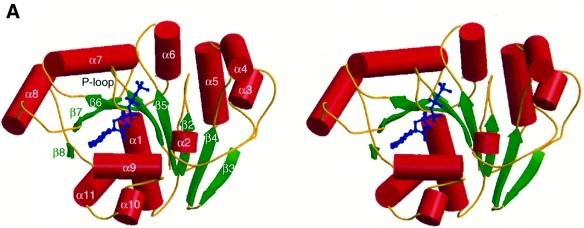
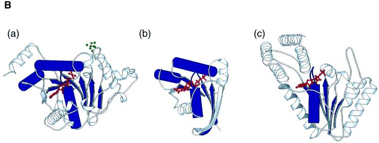
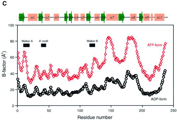
Fig. 1. Structure of P.furiosus MinD. (A) Stereo diagram of MinD in complex with AMPPCP. The helices are shown as red cylinders, β-strands as green arrows. The bound nucleotide is shown with thickened blue bonds. (B) Ribbon diagram of NIP (a), H-Ras (b) and NSF (c), shown in the same orientation as in (A). Structural elements compared with MinD are coloured in blue (see text). The bound nucleotides, ADP-AlF4– (a), GTP (b) and ATP (c) are shown in red; the 4Fe:4S of NIP is in green. (C) A plot of the average main chain temperature factors for MinD–nucleotide complexes (red line for the ATP form and black for the ADP form). Secondary structural elements are indicated on the top.

An official website of the United States government
Here's how you know
Official websites use .gov
A
.gov website belongs to an official
government organization in the United States.
Secure .gov websites use HTTPS
A lock (
) or https:// means you've safely
connected to the .gov website. Share sensitive
information only on official, secure websites.


