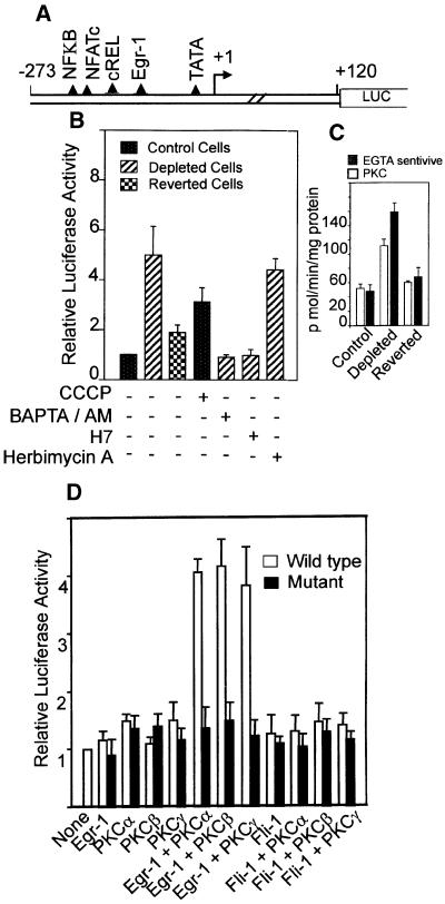
Fig. 5. Mechanism of activation of cathepsin L promoter by mitochondrial stress. (A) Structure of murine cathepsin L promoter and potential protein binding motifs based on nucleotide sequence. (B) Transcriptional activity of promoter construct in pGL3 reporter vector (Promega, Madison, WI). Details of transfection with reporter and renila luciferase as internal control and analysis of luciferase activity were as described in Materials and methods. Various agents as indicated were added to transfected cells also as described in Materials and methods. (C) PKC activity was assayed based on the level of inhibition with a PKC α, β and γ-specific pseudosubstrate inhibitor peptide. Ca2+-sensitive activity represents the level of inhibition with 5 mM EGTA. (D) Effects of coexpression with Egr-1 and PKC isoform-specific cDNAs on the transcriptional activity of the cathepsin L promoter in control C2C12 cells. Values represent the average ± SEM of 4–6 transfection assays in all cases.

An official website of the United States government
Here's how you know
Official websites use .gov
A
.gov website belongs to an official
government organization in the United States.
Secure .gov websites use HTTPS
A lock (
) or https:// means you've safely
connected to the .gov website. Share sensitive
information only on official, secure websites.
