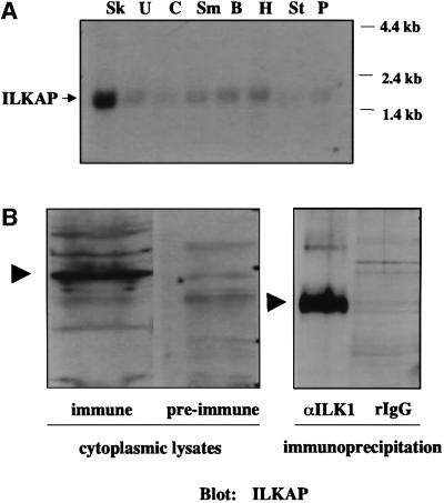
Fig. 2. ILKAP transcript is preferentially expressed in skeletal muscle. (A) A partial cDNA representing amino acid residues 269–392 of ILKAP was 32P labelled, and used to probe a northern blot of poly(A)+ RNAs from human striated and smooth muscle tissues. Sk, skeletal muscle; U, uterus; C, colon; Sm, small intestine; B, bladder; H, heart; St, stomach; P, prostate. (B) Left panel: a polyclonal antibody was raised to an ILKAP fusion protein (residues 269–392) and affinity purified over an immobilized ILKAP column. Pre-immune serum from the same rabbit was subjected to the same purification protocol, as a negative control. Cytoplasmic lysates of HEK 293 cells were analysed by western blotting using the ILKAP immune and pre-immune sera as indicated. Right panel: cytoplasmic 293 cell lysates were immunoprecipitated with rabbit IgG, or affinity-purified rabbit polyclonal ILK1 antibody, covalently coupled to CNBr-activated Sepharose beads. Unfractionated cytoplasmic lysates and ILK1 immunoprecipitates each indicated a single ILKAP band, migrating with an apparent mol. wt of 47 kDa. Immune complexes were analysed by western blotting, using affinity-purified ILKAP antibody. Arrowheads indicate p47ILKAP migration on 12% SDS–PAGE.

An official website of the United States government
Here's how you know
Official websites use .gov
A
.gov website belongs to an official
government organization in the United States.
Secure .gov websites use HTTPS
A lock (
) or https:// means you've safely
connected to the .gov website. Share sensitive
information only on official, secure websites.
