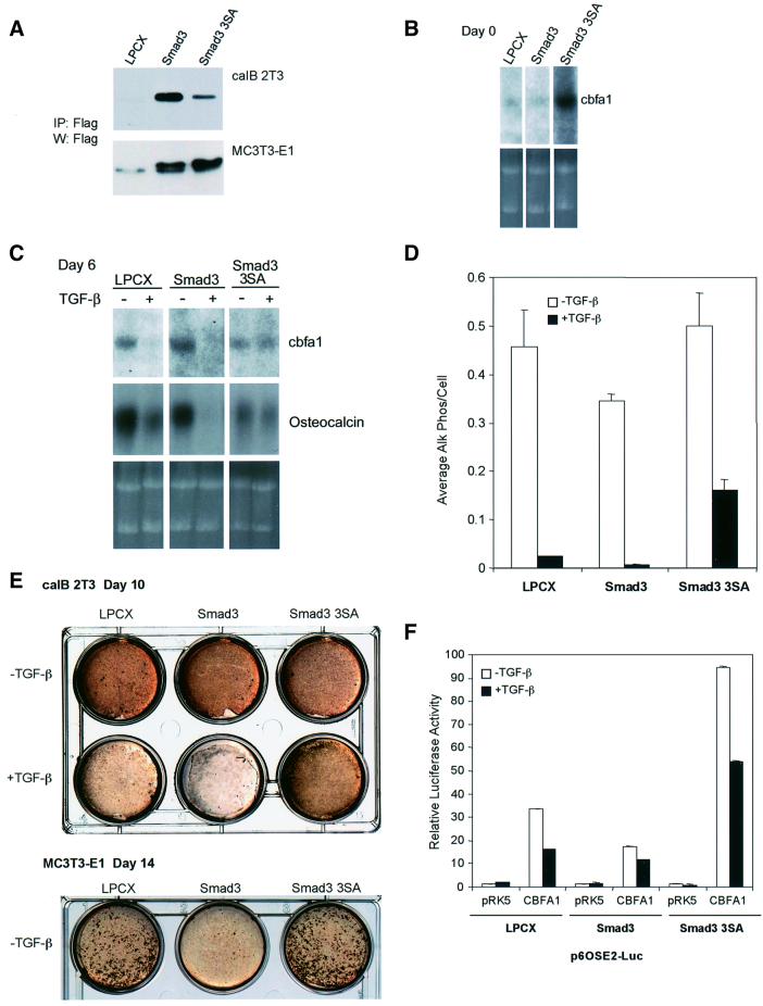
Fig. 9. Stable expression of wild-type or dominant-negative Smad3 in caIB 2T3 cells and MC3T3-E1 cells alters osteoblast differentiation. (A) Immunoprecipitation followed by western blot analysis demonstrates expression of Flag-tagged Smad3 or Smad3-3SA in cell lysates of stably infected caIB 2T3 and MC3T3-E1 cells. LPCX is the control empty vector. (B) Alterations in Smad3 signaling affect endogenous cbfa1 mRNA expression in caIB 2T3 cells at day 0, i.e. prior to exposure to differentiation conditions, in LPCX control cells, or cells stably expressing Smad3 or Smad3-3SA. The top panel shows cbfa1 northern hybridizations, while the lower panel shows the ethidium bromide-stained gel. (C) Alterations in Smad3 signaling affect endogenous cbfa1 and osteocalcin mRNA expression in differentiating caIB 2T3 cells at day 6 in differentiation conditions, in the absence or presence of TGF-β. The top panels show hybridizations for cbfa1 or osteocalcin mRNA using RNA from LPCX control cells or cells stably expressing Smad3 or Smad3-3SA, while the lower panel shows the ethidium bromide-stained gel. (D) Alterations in Smad3 signaling affect alkaline phosphatase activity in caIB 2T3 cells and the extent of inhibition by TGF-β (5 ng/ml). Cells were incubated in differentiation medium for 6 days in the presence or absence of TGF-β (5 ng/ml). Cell lysates were then assayed for alkaline phosphatase activity. Values are expressed per cell. (E) Alterations in Smad3 signaling affect matrix mineralization in caIB 2T3 and MC3T3-E1 cells and the extent of inhibition by TGF-β (5 ng/ml) in caIB 2T3 cells. Confluent cells were incubated for 10 (caIB 2T3 cells) or 14 days (MC3T3-E1 cells) in differentiation medium, in the presence or absence of TGF-β, and then stained for mineralization (brown) using the von Kossa method. (F) Alterations in Smad3 signaling affect the transactivation of the p6OSE2-reporter by CBFA1. Stably infected LPCX control or stable caIB 2T3 cells expressing Smad3 or Smad3-3SA were transiently transfected with the p6OSE2-Luc reporter plasmid, with or without an expression plasmid for CBFA1 (pRK5-CBFA1). Luciferase expression in the absence or presence of TGF-β (1 ng/ml) was scored as in Figure 2.

An official website of the United States government
Here's how you know
Official websites use .gov
A
.gov website belongs to an official
government organization in the United States.
Secure .gov websites use HTTPS
A lock (
) or https:// means you've safely
connected to the .gov website. Share sensitive
information only on official, secure websites.
