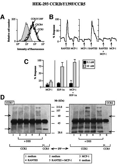
Fig. 2. The mutant CCR2bY139F receptor impairs the response to RANTES but to SDF-1α. (A) CCR2bY139F/CCR5 double-transfected HEK-293 cells were incubated with biotin-labeled mAbs to CCR2, CCR5 or CXCR4 or their respective isotype-matched control mAbs, followed by isothiocyanate-labeled streptavidin. (B) Ca2+ flux was triggered by 10 nM RANTES, 10 nM MCP-1, or a combination of both chemokines (0.1 nM each) as indicated using CCR2bY139F/CCR5-co-transfected HEK-293 cells. Results are expressed as a percentage of the maximum chemokine-induced Ca2+ response. The figure depicts one representative experiment of four performed. (C) Ca2+ mobilization was determined as in Figure 1B, following stimulation of CCR2b Y139F/CCR5-co-transfected HEK-293 cells with different concen trations of MCP-1 or SDF-1α, separately or simultaneously as indicated. Results are expressed as a percentage of the maximum chemokine-induced response. The mean ± SD of three independent experiments is shown. (D) HEK-293 cells co-transfected with the CCR2bY139F and CCR5 receptors were processed as in Figure 1D. Arrows indicate the monomer and the dimer.

An official website of the United States government
Here's how you know
Official websites use .gov
A
.gov website belongs to an official
government organization in the United States.
Secure .gov websites use HTTPS
A lock (
) or https:// means you've safely
connected to the .gov website. Share sensitive
information only on official, secure websites.
