
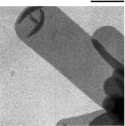
Fig. 5. Electron micrograph of a negatively stained two-dimensional crystal of SecYEG. Crystals stained by uranyl acetate were visualized using a Philips CM12 electron microscope. The tubular vesicle shown is flattened to the carbon surface and contains a two-dimensional crystal on each face. Bar, 0.5 µm.