
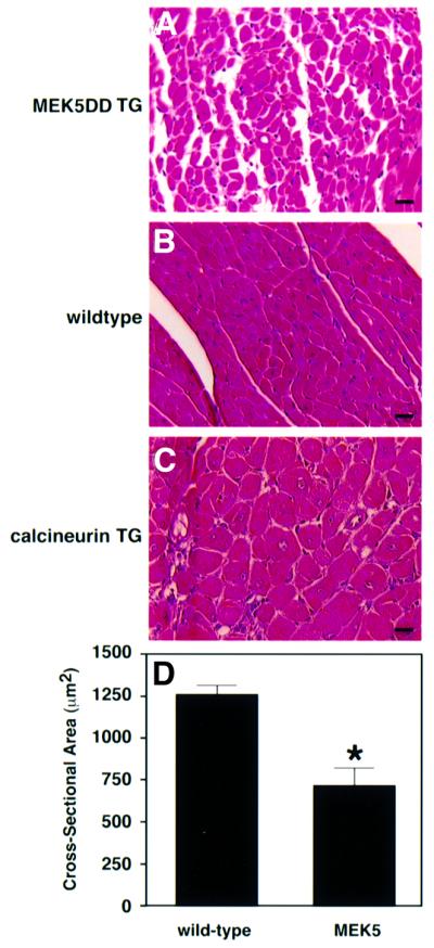
Fig. 8. MEK5DD transgenic hearts show reduced myofiber cross-sectional area relative to wild type. Hearts were removed from 8-week-old wild-type and from MEK5DD and calcineurin transgenic mice, fixed, sectioned and stained with hematoxylin–eosin. Dramatic differences in myocyte cross-sectional area are apparent in hematoxylin–eosin-stained sections from (A) MEK5DD transgenic hearts, (B) wild-type hearts and (C) calcineurin transgenic hearts. Bar, 20 µm. (D) The cross-sectional area of myocytes from 8-week-old wild-type and MEK5DD transgenic mice was quantitated using a computerized morphometric system. Measurements were made on equivalent sections from five wild-type and five transgenic hearts, and, within each section, measurements were taken from left and right ventricle, septum and papillary muscle (10 measurements each). The average result ± SD is shown; *P <0.001.