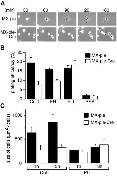
Fig. 4. Requirement for C3G in cell adhesion and cell spreading. (A) MEF-hC3G cells were infected with MX-pie-Cre or MX-pie. After 48 h, cells were trypsinized, kept in suspension for 1 h and plated on dishes coated with collagen type I. The cells were observed by time-lapse microscopy. We show representative photographs at the indicated time points. (B) MEF-hC3G cells were infected with MX-pie or MX-pie-Cre. After 48 h, cells were trypsinized and labeled with BCECF, AM. The cells were plated on 96-well black-colored plates coated with the reagents indicated at the bottom and incubated for 1 h at 37°C in a CO2 incubator. Cells were washed three times with HBSS, and the fluorescence intensity was measured at excitation and emission wavelengths of 488 and 530 nm, respectively. Plating efficiency is shown as the average for three wells, with the SE. (C) MEF-hC3G cells infected with MX-pie or MX-pie-Cre were plated to dishes coated with collagen (Col-I) or poly-l-lysine (PLL). Twenty EGFP-positive cells were photographed after 1 and 3 h and measured for size. Average and SE are shown.

An official website of the United States government
Here's how you know
Official websites use .gov
A
.gov website belongs to an official
government organization in the United States.
Secure .gov websites use HTTPS
A lock (
) or https:// means you've safely
connected to the .gov website. Share sensitive
information only on official, secure websites.
