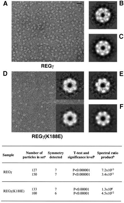
Fig. 2. Electron micrographs and averaged images of REGγ and REGγ (K188E) particles. Shown are fields of purified, negatively stained REGγ (A) and REGγ (K188E) (D), and the respective correlation averages of particles with 7-fold symmetry as judged by statistical analysis (B and E). (C and F) These images were explicitly symmetrized. (D, top inset) Correlation-average of image displaying 6-fold symmetry and (D, bottom inset) the corresponding symmetrized images. The averaged images (B), (E) and (D) (inset) represent 150, 133 and 100 particles, and have resolutions of 26, 28 and 28 Å, respectively. For REGγ (B and C), the inner ring of density is resolved into seven units whereas REGγ (K188E) (E and F) shows this feature as a continuous ring, presumably reflecting the lower resolution of the latter images. Bar, 40 nm (A) and 5 nm (B). Table: aindependent data sets from micrographs of different fields of the same grid in each sample; bthe spectral ratio product is shown only for the radial zones in which these symmetries were detected most strongly (at a radius of 5 nm).

An official website of the United States government
Here's how you know
Official websites use .gov
A
.gov website belongs to an official
government organization in the United States.
Secure .gov websites use HTTPS
A lock (
) or https:// means you've safely
connected to the .gov website. Share sensitive
information only on official, secure websites.
