
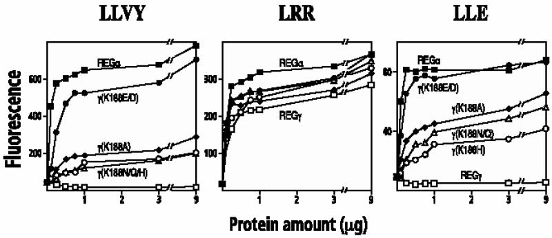
Fig. 3. Activation of fluorogenic peptide hydrolysis by REGγ Lys188 mutants. Human red blood cell proteasomes (170 ng) were mixed with increasing amounts of purified REG variants. The reaction was started by adding 50 µl of 200 µM Suc-LLVY-MCA (left), Boc-LRR-MCA (middle) or Boc-LLE-βNA (right) in 10 mM Tris pH 7.5. After a 10 min incubation, the reaction was quenched with 200 µl of cold 100% ethanol, and the released MCA or βNA was measured fluorometrically. Each data point represents the mean of three measurements from a single experiment; equivalent results were observed in at least two experiments using different preparations of the various REG proteins. Symbol representation: REGα, filled squares; REGγ, open squares; REGγ (K188E) or REGγ (K188D), filled circles; REGγ (K188H), open circles; REGγ (K188Q) or REGγ (K188N), open triangles; REGγ (K188A), filled diamonds. Data from REGγ (K188D) and REGγ (K188E) are combined since their activities are indistinguishable. The same is true for REGγ (K188N) and REGγ (K188Q).