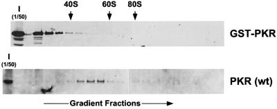
Fig. 3. Ribosome association of wild-type PKR, but not the GST–PKR fusion protein. Transformants of strain J82 (expressing eIF2α-S51A) containing the PKR plasmid p1420 or the GST–PKR plasmid pC661 were grown in SGal medium to an OD600 ∼1.5. Whole-cell extracts were prepared in the presence of cycloheximide (50 mg/ml) and MgCl2 (10 mM), and then subjected to velocity sedimentation on 5–47% sucrose gradients as described previously (Zhu et al., 1997; Romano et al., 1998b). The gradients were fractionated while monitoring absorbance at 254 nm to identify the positions of free 40 and 60S subunits, and 80S monosomes (as indicated by the arrows). The distribution of PKR and GST–PKR along the gradients was visualized by SDS–PAGE and immunoblot analysis using polyclonal antiserum raised against the GST–PKR fusion protein. The first lane in each panel was loaded with 1/50 of the input (I) extracts fractionated on the gradients.

An official website of the United States government
Here's how you know
Official websites use .gov
A
.gov website belongs to an official
government organization in the United States.
Secure .gov websites use HTTPS
A lock (
) or https:// means you've safely
connected to the .gov website. Share sensitive
information only on official, secure websites.
