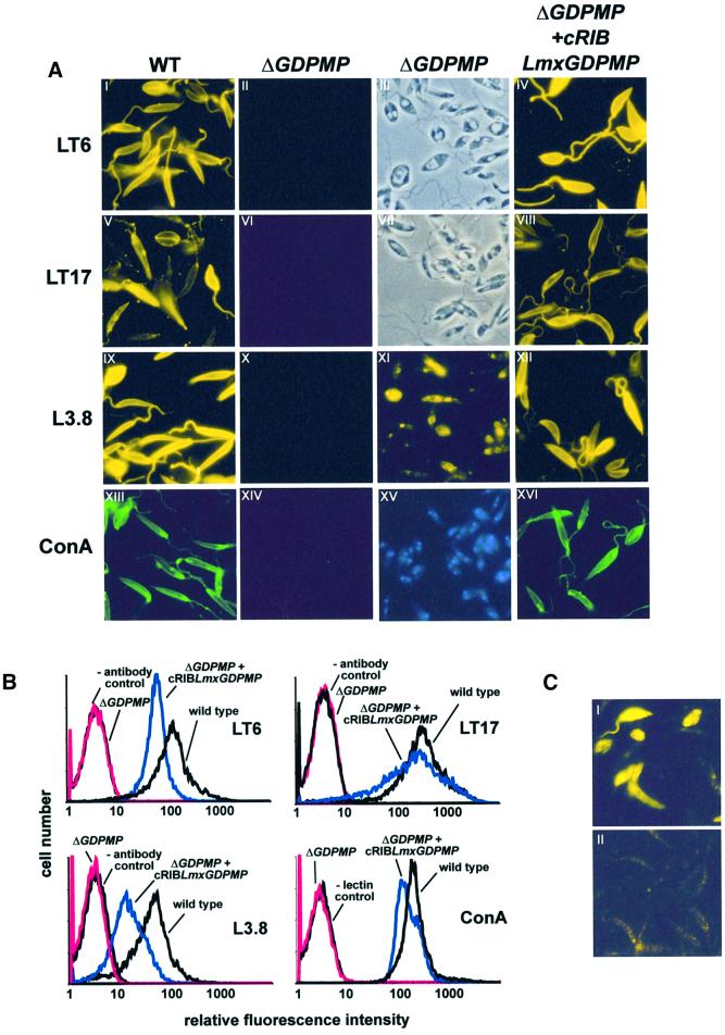
Fig. 5. Immuno-/lectin fluorescence microscopy and FACS analysis of Leishmania WT and mutant promastigotes. (A) Immuno-/lectin fluorescence microscopy of fixed L.mexicana promastigotes. The cells were not permeabilized, except for XI. The parasite lines and the mAbs/lectins used are indicated by the labeling of columns and rows. Exposure times within rows (A) are identical, except for XI, which is ∼20× overexposed compared with IX. III and VII in (A) are phase-contrast light microscopy images of II and VI, respectively. (B) FACS analysis of live L.mexicana promastigotes. The parasite lines and the mAbs/lectins used are indicated in each panel. (C) IFM of saponin-permeabilized L.mexicana WT promastigotes labeled with rabbit anti-GDPMP serum (1:200) (I) and pre-immune serum (1:200) (II). Exposure times were identical in I and II.

An official website of the United States government
Here's how you know
Official websites use .gov
A
.gov website belongs to an official
government organization in the United States.
Secure .gov websites use HTTPS
A lock (
) or https:// means you've safely
connected to the .gov website. Share sensitive
information only on official, secure websites.
