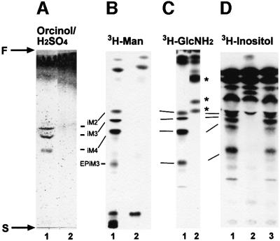
Fig. 6. Silica gel 60 HPTLC analysis of the predominant promastigote glycolipids of L.mexicana in WT (lane 1), ΔGDPMP (lane 2) and ΔGDPMP + cRIBLmxGDPMP (lane 3) parasites. (A) Total lipids from 2 × 10⁸ promastigotes visualized by orcinol/H2SO4 spraying. (B) Fluorography of total lipids from 2.5 × 10⁷ [3H]Man-labeled promastigotes (∼100 000 c.p.m.). (C) Fluorography of total lipids from 5 × 10⁶ [3H]GlcNH2-labeled promastigotes (∼100 000 c.p.m.). (D) Fluorography of total lipids from 5 × 10⁶ [3H]myo-inositol-labeled promastigotes (∼100 000 c.p.m.). The positions of the abundant L.mexicana GIPLs (McConville et al., 1993) are indicated by the bars, and the start and front of the TLCs are marked by S and F, respectively. Asterisks mark new [3H]GlcNH2-labeled compounds accumulating in the ΔGDPMP mutant.

An official website of the United States government
Here's how you know
Official websites use .gov
A
.gov website belongs to an official
government organization in the United States.
Secure .gov websites use HTTPS
A lock (
) or https:// means you've safely
connected to the .gov website. Share sensitive
information only on official, secure websites.
