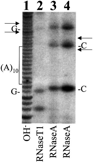
Fig. 5. Enzymatic sequencing of the T7 transcript containing codons 8–14 of the HCV sequence. Lane 1, partial alkaline (OH–) hydrolysis of the HCV RNA; lane 2, partial digestion with RNase T1, which cuts after G; lanes 3 and 4, partial digestion with RNase A, which cuts after U and C. The locations of the 10 A stretch and its two flanking C residues and G residues are indicated. Arrows indicate the locations of RNase T1 and RNase A bands that would be produced if there was a one nucleotide deletion or a two nucleotide insertion in the 10 A stretch. Note that there was no apparent sequence heterogeneity flanking the 10 A stretch.
