
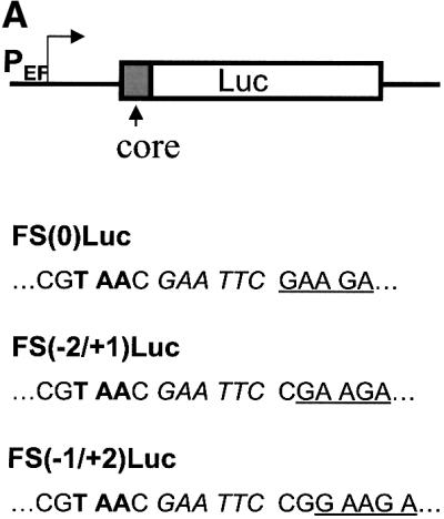
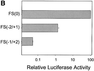
Fig. 6. Analysis of the ribosomal frameshift efficiency in Huh7 cells. (A) Schematic illustration of the luciferase reporter constructs. PEF indicates the EF1α promoter, Luc indicates the luciferase reporter, and core indicates codons 1–14 of the core protein sequence. Sequences located at the fusion junction of the core protein and the luciferase reporter (underlined) are also shown. Italic letters indicate the EcoRI site that was used to fuse the two sequences. Bold letters denote the termination codon located in the –1/+2 reading frame. (B) The luciferase reporter assay. Huh7 cells were transfected with various luciferase reporter constructs, and the relative frameshift efficiencies were determined by the procedures described in Materials and methods. The experiments were carried out in triplicate and repeated at least twice. Data represent the average of the results.