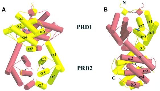
Fig. 3. (A) Ribbon drawing using the Molscript package of the LicT-PRD dimer perpendicular to the 2-fold symmetry axis (Carson, 1991; Kraulis, 1991). The individual monomers are coloured yellow and pink. The subdomains PRD1 and PRD2 and their individual α-helices (α1–α5) are indicated. The phosphorylatable histidines (His100 and His159) in PRD1 and the mutated aspartates in PRD2 (Glu207 and Glu269) are shown in ball-and-stick representation. (B) The LicT-PRD dimer viewed in the same direction to that shown in (A) but rotated over 90° along the 2-fold symmetry axis.

An official website of the United States government
Here's how you know
Official websites use .gov
A
.gov website belongs to an official
government organization in the United States.
Secure .gov websites use HTTPS
A lock (
) or https:// means you've safely
connected to the .gov website. Share sensitive
information only on official, secure websites.
