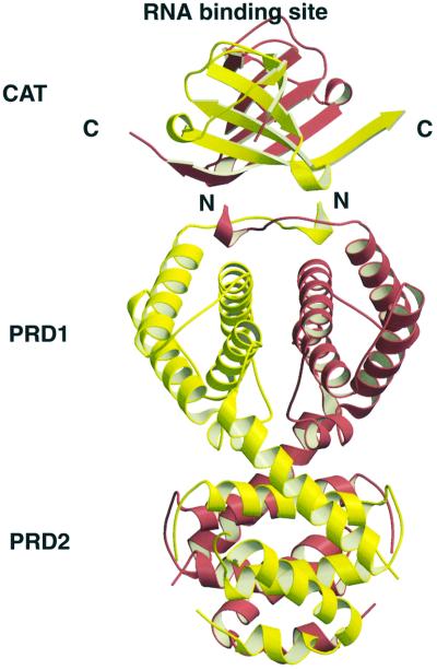
Fig. 7. Ribbon presentation of the model of the intact LicT protein using the coordinates of the crystal structures of the CAT domain (Declerck et al., 1999) and the PRD (this study). The individual monomers are in yellow and red. The N-terminus (N) of the PRD and the C-terminus (C) of the CAT domain are indicated. The model was created by first aligning the 2-fold axes dimer of the CAT and PRD domains and then approaching at best the C-terminus of CAT and the N-terminus of PRD. The RNA-binding surface as determined by NMR footprinting and site-directed mutagenesis is also indicated (Manival et al., 1997).

An official website of the United States government
Here's how you know
Official websites use .gov
A
.gov website belongs to an official
government organization in the United States.
Secure .gov websites use HTTPS
A lock (
) or https:// means you've safely
connected to the .gov website. Share sensitive
information only on official, secure websites.
