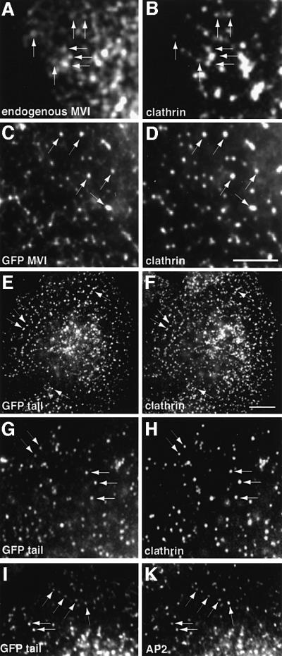
Fig. 4. Co-localization of myosin VI with AP-2 and clathrin in NRK cells. (A) An untransfected cell stained for endogenous myosin VI with an antibody to the whole tail. Endogenous myosin VI not containing the large insert (A) shows partial co-localization with clathrin (B) in NRK cells. NRK cells transiently expressing whole myosin VI from chicken brush border containing the large insert (C) or only the tail containing the large insert, both tagged with GFP (E, G and I), were stained with antibodies to AP-2 (K) or clathrin (D, F and H). The GFP–myosin VI and the tail show co-localization with clathrin and AP-2 at the plasma membrane. Bar: 20 µm.

An official website of the United States government
Here's how you know
Official websites use .gov
A
.gov website belongs to an official
government organization in the United States.
Secure .gov websites use HTTPS
A lock (
) or https:// means you've safely
connected to the .gov website. Share sensitive
information only on official, secure websites.
