
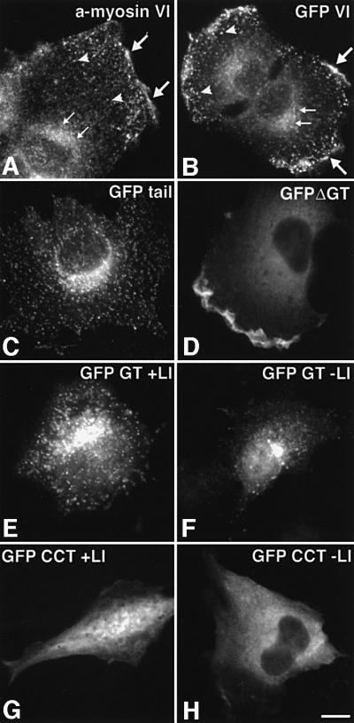
Fig. 6. Localization of GFP–myosin VI and GFP–myosin VI deletion mutants in NRK cells. (A) An untransfected cell stained for endo genous myosin VI with an antibody to the whole tail (a-myosin VI). Myosin VI is enriched in the perinuclear area (small arrows), in ruffles at the edge of the cell (large arrows) and in a vesicular staining pattern in the thin leading lamella (arrowheads). (B–H) NRK cells transiently expressing GFP-tagged constructs: (B) whole myosin VI tagged with GFP [symbols as in (A)]; (C) GFP–tail; (D) GFP–myosin VI without the GT (GFP ΔGT); (E) the GT containing the large insert (GFP GT + LI); (F) the GT without the large insert (GFP GT – LI); (G) the coiled-coil domain of the tail tagged with GFP containing the large insert (GFP CCT + LI); and (H) the coiled-coil domain without the large insert (GFP CCT – LI). The GT together with the large insert are important for targeting to vesicular structures [see (B), (C), (E) and (F) compared with (D), (G) and (H)]. Only expression constructs including the motor domain were found in ruffles [compare (B) and (D) with (C), (E) and (F)]. Bar: 15 µm.