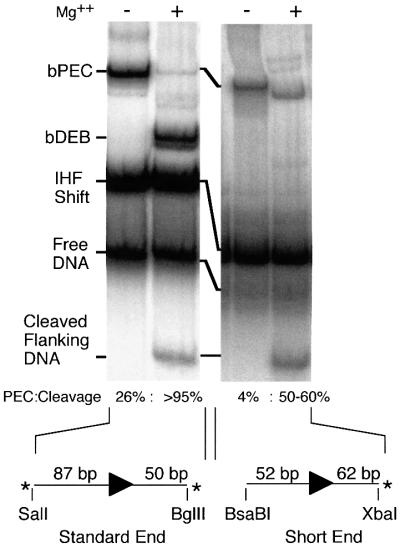
Fig. 6. Disruption of the subterminal contacts using a short transposon end fragment. The standard gel shift assay was performed using the standard BglII–SalI transposon end fragment or a short fragment with only 52 bp of transposon DNA. Assignment of the bands is as in Figure 1B. With the standard fragment, the quantity of bDEB appears greater than the cleaved flanking DNA because the SalI site is labeled more efficiently than the BglII site. The short end is only labeled on the flanking DNA because the opposite end is blunt.

An official website of the United States government
Here's how you know
Official websites use .gov
A
.gov website belongs to an official
government organization in the United States.
Secure .gov websites use HTTPS
A lock (
) or https:// means you've safely
connected to the .gov website. Share sensitive
information only on official, secure websites.
