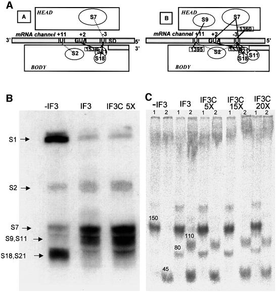
Fig. 5. Effect of IF3 and IF3C on the mRNA shift on the 30S ribosomal subunit. (A) To illustrate the IF3-induced repositioning of the mRNA on the 30S subunit described previously (La Teana et al., 1995), we show the main site-directed mRNA–30S cross-linking sites obtained in the absence (left) and presence (right) of IF3. (B) Autoradiography of the SDS–PAGE separation of the ribosomal proteins (indicated by the arrows on the left side) cross-linked to the mRNA in initiation complexes formed, as indicated above the corresponding lanes, in the absence or presence of either IF3 or IF3C (at a 5-fold higher concentration than IF3). (C) Autoradiography of RNaseH digestion products of 16S rRNA UV cross-linked to 32P-labelled mRNA within 30S–mRNA complexes prepared in the presence of IF1, IF2 and, where indicated, IF3 or increasing amounts of IF3C. The RNaseH digestion was carried out in the presence of oligonucleotides complementary to three regions of 16S rRNA, namely (A) 1298–1307, (B) 1382–1391 and (C) 1491–1507. Two separate digestions were performed with two different pairs of oligonucleotides, A/B (lanes marked 1) and B/C (lanes marked 2), respectively. The bands marked 45 and 150, which are obtained with all types of complexes, correspond to cross-links between –3 and +2 of mRNA and a single site (1530) of 16S rRNA, while the two additional bands marked 80 and 110, obtained exclusively from the complexes containing IF3 or IF3C, correspond to cross-links between –3 of the mRNA and 1360 of 16S rRNA, and between +11 of the mRNA and 1395 of 16S rRNA, respectively. The experimental details are identical to those described previously and it should be noted that although the electrophoretic analysis does not allow the separation of S9/S11 and S18/S21, all these proteins were demonstrated to be cross-linked to the mRNA by immunological analysis (Dontsova et al., 1991; La Teana et al., 1995).

An official website of the United States government
Here's how you know
Official websites use .gov
A
.gov website belongs to an official
government organization in the United States.
Secure .gov websites use HTTPS
A lock (
) or https:// means you've safely
connected to the .gov website. Share sensitive
information only on official, secure websites.
