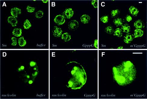
Fig. 5. Treatment with the m7GpppG cap analog does not alter the subnuclear distribution of Sm speckles or nucleoli. Cells were treated with buffer, GpppG or m7GpppG as indicated. U937 and K562 cells were stained for Sm and nucleolin, respectively. The objective is 100×. Scale bars = 5 µM. Confocal micrographs represent single sections through the plane of the cells.

An official website of the United States government
Here's how you know
Official websites use .gov
A
.gov website belongs to an official
government organization in the United States.
Secure .gov websites use HTTPS
A lock (
) or https:// means you've safely
connected to the .gov website. Share sensitive
information only on official, secure websites.
