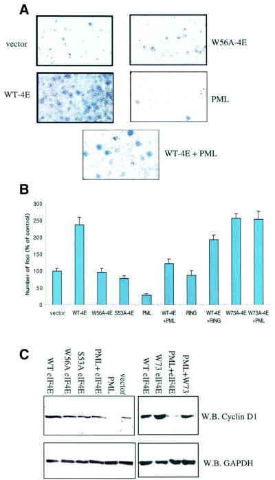
Fig. 7. Growth suppression and Cyclin D1 levels in PML–/– cells. (A) Anchorage-dependent foci formation. Foci stained with Giemsa formed as a result of different transfections. Full-length PML and eIF4E were used in these assays. Cells were transfected as indicated: wild-type eIF4E (WT-4E), PML, PML site I RING mutant (RING), cap-binding mutant of eIF4E (W56A-4E), PML-binding mutant of eIF4E (W73A-4E), PML and WT-4E (WT-4E + PML) and vector. Identically sized areas were taken from representative regions of each petri dish. These results were quantitated in (B). Foci were counted in seven dishes per treatment, and the values represent the mean ± SD. Results are the average of at least two independent experiments. (C) Cyclin D1 levels. Western analysis of the indicated experiments 24 h after transfection. Fifty micrograms of total protein were loaded into each lane. GAPDH and Cyclin D1 proteins were detected using western analysis.

An official website of the United States government
Here's how you know
Official websites use .gov
A
.gov website belongs to an official
government organization in the United States.
Secure .gov websites use HTTPS
A lock (
) or https:// means you've safely
connected to the .gov website. Share sensitive
information only on official, secure websites.
