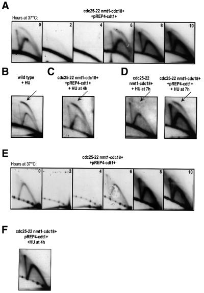
Fig. 6. Origin firing in response to Cdt1. (A) 2D gels of genomic DNA extracted from cdc25-22 nmt1-cdc18+ pREP4-cdt1+ cells probed for ars3001. Samples were collected every 2 h following the shift to 37°C in the absence of thiamine. (B) 2D gel of wild-type cells treated with HU (11 mM) for 3.5 h at 32°C, probed for ars3001. The arrow indicates the stronger bubble arc. (C) 2D gel of cdc25-22 nmt1-cdc18+ pREP4-cdt1+ cells shifted to 37°C in the absence of thiamine and treated with HU (24 mM) at 4 h. Samples were collected at 6 h at 37°C, after 2 h of treatment with HU. Gels were probed for ars3001. (D) 2D gels of cdc25-22 nmt1-cdc18+ pREP4 and cdc25-22 nmt1-cdc18+ pREP4-cdt1+ cells shifted to 37°C in the absence of thiamine, treated with HU (24 mM) at 7 h and collected 1 h later. Gels were probed for ars3001. (E) 2D gel of genomic DNA extracted from cdc25-22 nmt1-cdc18+ pREP4-cdt1+ cells and probed for a non-origin region downstream of ars3001. Samples were collected every 2 h following the shift to 37°C in the absence of thiamine. (F) 2D gel of cdc25-22 nmt1-cdc18+ pREP4-cdt1+ cells shifted to 37°C in the absence of thiamine and treated with HU (24 mM) at 4 h. Cells were collected at 10 h after 6 h of treatment with HU. The 2D gel was probed with the same fragment as described in (E).

An official website of the United States government
Here's how you know
Official websites use .gov
A
.gov website belongs to an official
government organization in the United States.
Secure .gov websites use HTTPS
A lock (
) or https:// means you've safely
connected to the .gov website. Share sensitive
information only on official, secure websites.
