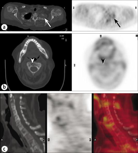
Figure 11.
A patient with low-grade lymphoma. (a) PET and CT images through a left apical mass with rib involvement show only mild FDG uptake (arrows), very unusual for lymphoma. (b) A CT image through C2 clearly shows a destructive lesion (arrowhead), but a PET image shows minimal FDG uptake (arrowhead). (c) Sagittal CT, PET, and fusion images of the cervical spine show minimal or no FDG uptake in the extensive lytic disease seen on CT.

