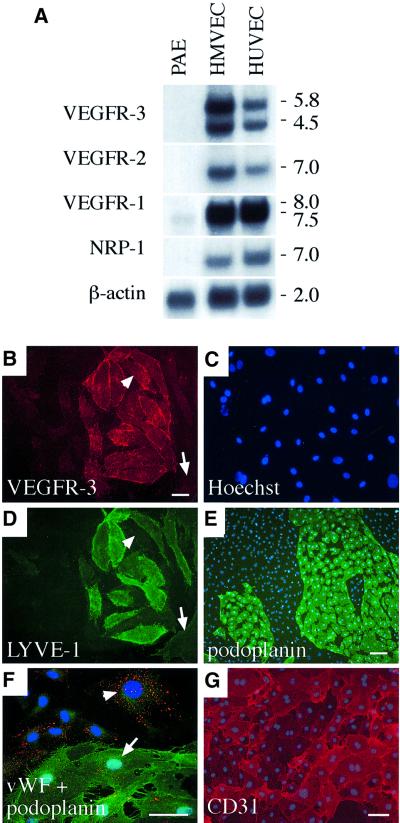
Fig. 1. (A) Expression of VEGFR mRNAs in primary human dermal microvascular endothelial cells (HMVECs), human umbilical vein endothelial cells (HUVECs) and in the porcine aortic endothelial (PAE) cell line. A northern blot containing 8 µg of the mRNAs was probed with radiolabelled cDNA fragments of human VEGF receptors and with β-actin for the control of equal loading. Numbers to the right denote the sizes of the transcripts (kb). (B–G) HMVECs consist of two distinct populations of blood vascular and lymphatic endothelial cells. Immunofluorescence double staining using antibodies against VEGFR-3 (B) and LYVE-1 (D) with conterstaining of the nuclei by Hoechst fluorochrome (C). Note that LYVE-1 expression is not detected in all VEGFR-3-positive cells (arrowhead in B and D) while some VEGFR-3-negative cells are also stained weakly with LYVE-1 antibodies (arrow in B and D). Immunolabelling with antibodies against podoplanin (green in E and F), vWF (red, F) and CD31 (G). The nuclei were stained with the Hoechst fluorochrome (E–G). vWF expression occurs primarily in the podoplanin-negative cells (arrowhead in F), but weaker expression is also detected on podoplanin-positive cells (arrow in F). Stainings in (B–E) were carried out using live cells on ice, and in (F) and (G) after PFA fixation. Scale bars in (B–D), (F) and (G), 50 µm; in (E), 100 µm.

An official website of the United States government
Here's how you know
Official websites use .gov
A
.gov website belongs to an official
government organization in the United States.
Secure .gov websites use HTTPS
A lock (
) or https:// means you've safely
connected to the .gov website. Share sensitive
information only on official, secure websites.
