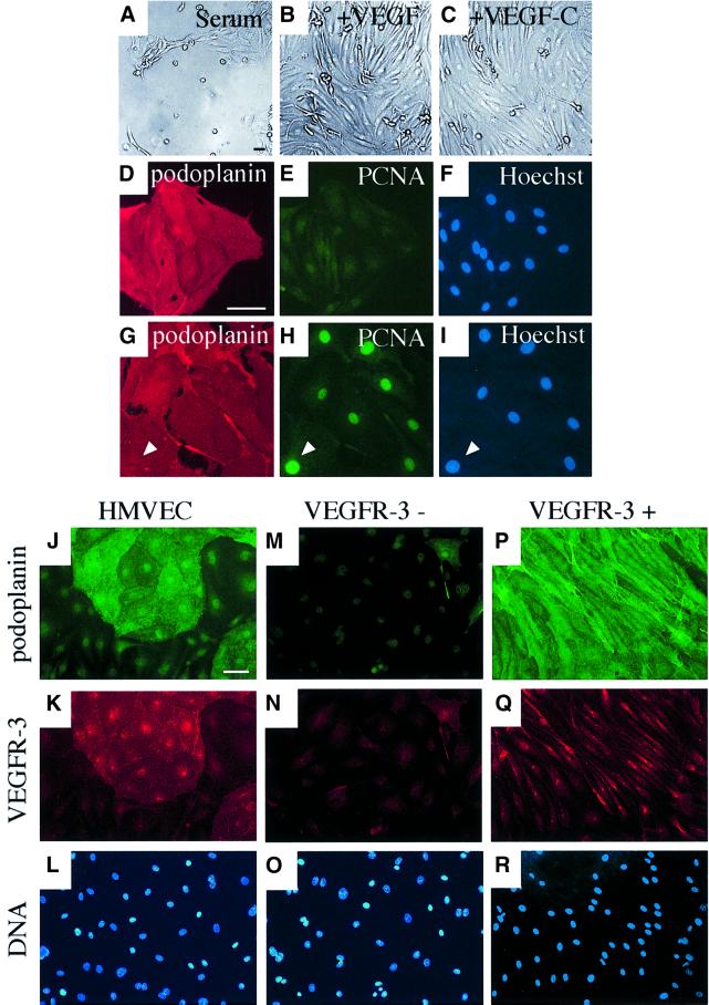
Fig. 4. Isolation of VEGFR-3/podoplanin-positive and -negative endothelial cells using magnetic microbeads. (A–I) Culture of VEGFR-3-expressing lymphatic endothelial cells in complete medium containing 5% serum (A) and supplemented with VEGF (10 ng/ml, B) or VEGF-C (100 ng/ml, C). Staining of the VEGFR-3-positive cells grown for 5 days after sorting in serum (D–F) or supplemented with VEGF-C (G–I) for podoplanin (red; D and G) or PCNA (green; E and H). The nuclei were stained with the Hoechst fluorochrome (F and I). Note that if supplemented with VEGF-C, the cells are stained for PCNA (arrowhead in G–I). Immunofluorescence double-staining of non-sorted cells (J–L) or VEGFR-3-negative (M–O) and VEGFR-3-positive (P–R) cell populations with antibodies against podoplanin (green; J, M and P) or VEGFR-3 (red; K, N and Q). The nuclei were stained with the Hoechst fluorochrome (L, O and R). The VEGFR-3+ cells were cultured in the presence of VEGF-C (100 ng/ml). Scale bars, 50 µm.

An official website of the United States government
Here's how you know
Official websites use .gov
A
.gov website belongs to an official
government organization in the United States.
Secure .gov websites use HTTPS
A lock (
) or https:// means you've safely
connected to the .gov website. Share sensitive
information only on official, secure websites.
