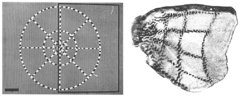
Fig. 2.

Retinotopic map of visual cortex in a macaque monkey. Left panel shows the visual stimulus with check-filled radial lines and concentric rings. Right panel shows the flattened visual cortex with representation of the radial lines and concentric rings marked by 2-DG. (Reproduced by permission from Tootell R.B.H., Switkes E., Silverman M.S. & Hamilton S.L. “Functional anatomy of macaque striate cortex. II. Retinotopic organization” in the Journal of Neuroscience 8(5), p. 1534 (Fig. 1) and p. 1535 (Fig. 2B); © 1988 by the Society for Neuroscience)
