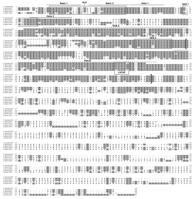
Fig. 1. Alignment of AHR amino acid sequences.

Deduced amino acid sequences of from Xenopus laevis AHR1α and AHR1β, Xenopus tropicalis AHR1, and Fundulus heteroclitus AHR1 and AHR2 (Karchner et al. 1999) were aligned using Clustal X, version 1.8 (Thompson et al. 1997). Identities are boxed a shaded. Dashes indicate gaps in alignment. Several putative functional domains are indicated: Basic and Helix-loop-helix domains are denoted with solid lines above the sequence: PAS A and PAS B domains with hatched lines, and the ligand binding domain with brackets. (Coumailleau et al. 1995; Fukunaga et al. 1995). NLS, nuclear localization signal (Ikuta et al. 1998), NES1, nuclear export signal 1 (Ikuta et al. 1998), NES2, nuclear export signal 2 (Berg and Pongratz 2001), and LXCXE motif, a retinoblastoma protein binding motif (Elferink et al. 2001; Puga et al. 2000a) are indicated with boxes.
