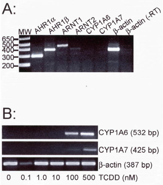
Figure 8. TCDD responsiveness in A6 cells.

X. laevis A6 cells were grown to near confluence and treated with DMSO or graded concentrations of TCDD. CYP1A6 and CYP1A7 mRNA expression was detected by RT-PCR with total RNA. (A) Expression of mRNAs encoding components of the AHR signaling pathway. Target transcripts are indicated above each lane. Mobility of DNA size makers (MW) are indicated at left. One β-actin reaction omitted the addition of reverse transcriptase during the cDNA synthesis phase (-RT). (B) Cyp1A inducibility. Cells treated for 24 hr with graded concentrations of TCDD (indicated at bottom). Total RNA was isolated and subjected to RT-PCR with primers specific for CYP1A6 and CYP1A7. Controls lacking reverse transcriptase (not shown) were performed for each concentration and primer pair.
