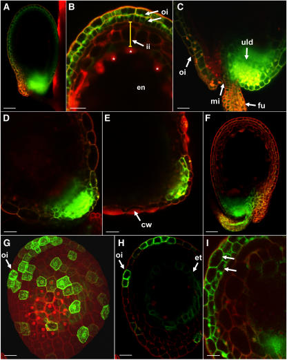
Figure 1.
Analysis of the symplastic connectivity and of post-phloem GFP movement in the outer integument of Arabidopsis seeds. A to E, Seeds from AtSUC2 promoter/GFP plants. A, Overview of the GFP fluorescence in a seed from the fifth silique (2 d old; globular stage of the embryo; bar = 50 μm). B, Magnification of the tip region of a seed similar to that shown in A. GFP fluorescence is detected in both cell layers of the outer integument (oi). No GFP fluorescence is seen in any of the three cell layers of the inner integument (ii; yellow bar) or in the endosperm (en). Some of the peripheral endosperm nuclei are marked with asterisks (bar = 25 μm). C, Magnification of the basal unloading (uld) domain at the end of the funiculus (fu) of a seed similar to that shown in A. Weak GFP fluorescence is seen in both cell layers of the outer integument (oi; mi, micropyle; bar = 25 μm). D, GFP fluorescence in a seed from the twelfth silique (6 d old; upturned-U stage of the embryo). At this stage, GFP is still unloaded from the funicular vascular bundle, but post-phloem movement of GFP in the outer integument is no longer detected (bar = 25 μm). E, GFP fluorescence in a seed from the fifteenth silique (10 d old; mature embryo). GFP fluorescence is seen only in few cells of the unloading domain. At this stage, cell wall (cw) thickening and development of the testa have already started (bar = 25 μm). F, Seed from an AtSUC2 promoter/GFP-sporamin plant. GFP-sporamin is unloaded from the vascular bundle at the end of the funiculus, but post-phloem movement in the outer integument is not detected (2 d old; globular stage of the embryo; bar = 40 μm). G and H, Seed from an AtGL2 promoter/tmGFP9 plant. G, View of the seed from the outside showing AtGL2 promoter activity in individual cells of the outer integument (oi; bar = 20 μm). H, Optical section through the seed shown in G confirming the AtGL2 promoter activity in the outermost cell layer of the outer integument (oi). A weak activity of the AtGL2 promoter is also seen in the innermost layer of the inner integument, the endothelium (et; bar = 20 μm). I, Seed of an AtGL2 promoter/GFP plant. GFP fluorescence is seen in both layers of the outer integument (arrows) showing that GFP can move cell to cell not only within the outer cell layer but also from the outer into the inner cell layer (bar = 25 μm). All images represent CLSM images. G is a maximum projection of a picture stack. All others represent optical sections. Red color results from propidium iodide staining of cell walls.

