
Fig. 10.
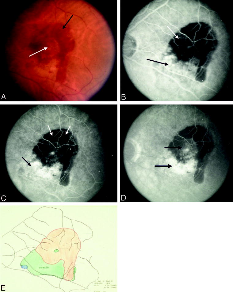
Subfoveal blood with or without choroidal neovascularization (CNV), Group B. A, Color photograph shows extensive subretinal hemorrhage (black arrow) with a clear zone centrally (white arrow). B, Early phase of the angiogram shows blocked fluorescence from blood (white arrow) with mild speckled fluorescence at the inferior border (black arrow). C, By the mid phase of the angiogram, the occult CNV is evident both at the inferior border (long black arrow) and within the central area of blocked fluorescence from sub-retinal hemorrhage (short black arrow). D, Leakage progresses into the late phase. E, Composite drawing using multiple stereoscopic photographs of angiograms shows interpretation of the boundaries of the lesion components.