
Fig. 9.
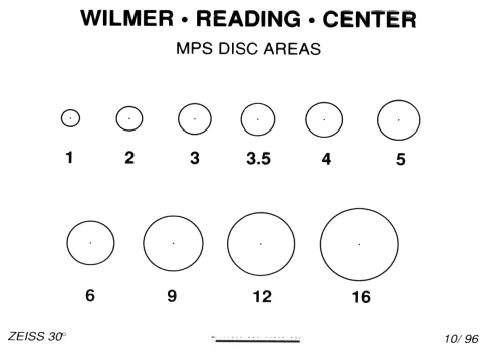
Disk area template. Template of clear acetate is placed on a 35-mm frame of the angiogram (made using a 30° fundus camera) to determine the lesion area. One Macular Photocoagulation Study (MPS) disk area is defined as an area of 2.54 mm2, assuming a 2.5 magnification of the image on the film relative to the size of the lesion on the retina. Thus, the 2 MPS disk area circle on the template has an area that covers 5.06 mm2 on the retina.