
Figure 3.
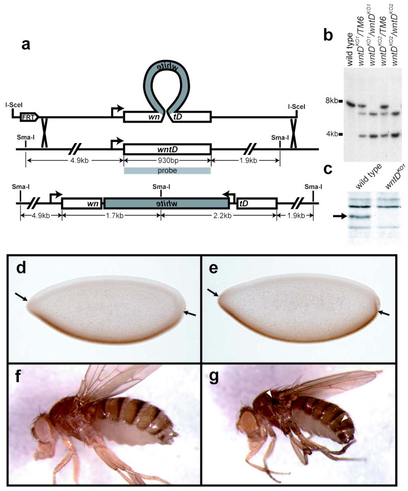
wntD knockout flies exhibit ectopic Dorsal activation. a, “Ends-out” knockout targeting scheme, illustrating how a white mini-gene was used to interrupt the wntD open reading frame. b, Southern blot of Sma-I digested genomic DNA, confirming proper integration of targeting construct. c, Anti-WntD Western blot of lysate from wild type and wntDKO1 embryos (arrow indicates size of WntD protein). d,e, yw (d) and yw; wntDKO1 (e) embryos stained with antibodies against Dorsal. Arrows show point of ventral-most nuclear Dorsal seen in control embryos. f,g adult female yw (f) and yw; wntDKO1 (g) flies. Arrowheads mark sites of ectopic melanization.