
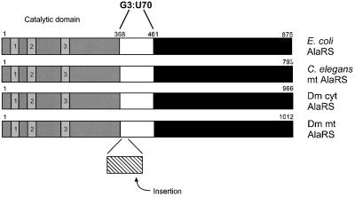
Fig. 8. Insertion of a 28 amino acid segment into Dm mt AlaRS. AlaRSs from E.coli, C.elegans mitochondria, D.melanogaster cytoplasm and D.melanogaster mitochondria are compared. The active site-containing domain of AlaRS with the three characteristic sequence motifs (1–3) of class II enzymes is shaded. The domain of E.coli AlaRS thought to contact the G3:U70 base pair (E.coli numbering shown) and homologous domains are white, while the insertion present in Dm mt AlaRS is shown with cross hatches. The C-terminal portions of the proteins are in black.