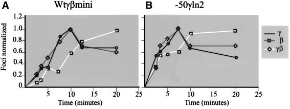
Fig. 5. Kinetics of reappearance of the single (γ or β) versus double (γβ) primary transcription signals after release of the DRB block to transcription elongation (see Gribnau et al., 1998). Transcription foci were counted in 12 day fetal liver nuclei after in situ hybridization with γ- and β-globin intron probes in nuclei fixed at different times after the release of the DRB block. The number of black (γ), gray (β) or white (γ + β) signals was plotted against time of fixation after the release of the block. (A) Signals observed in mice containing a single copy of the wtγβmini locus. (B) Signals from the line –50γln2. Symbols corresponding to the γ, β or γβ foci are shown on the right.

An official website of the United States government
Here's how you know
Official websites use .gov
A
.gov website belongs to an official
government organization in the United States.
Secure .gov websites use HTTPS
A lock (
) or https:// means you've safely
connected to the .gov website. Share sensitive
information only on official, secure websites.
