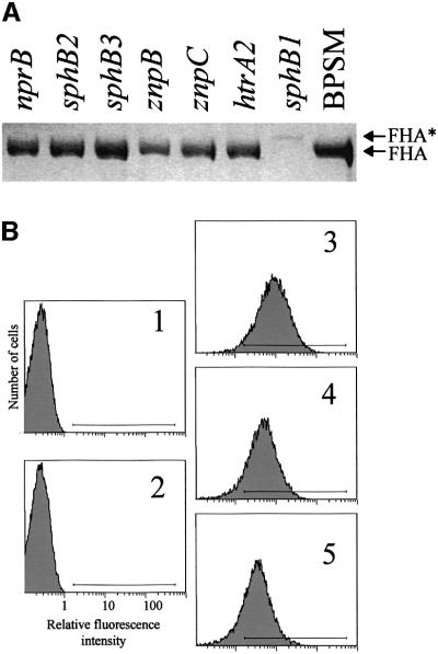
Fig. 1. FHA secretion and cell surface exposure in B.pertussis parental and protease mutant strains. (A) Identical volumes (20 µl) of non-concentrated supernatants from each culture at late logarithmic phase of growth were analysed by SDS–PAGE. The gel was stained with Coomassie Blue. BPSM is the parental strain, and each derived strain is denoted by its genotype relative to BPSM. FHA and FHA* represent the positions of mature FHA and a slightly larger derivative, respectively. (B) BPGR4 (fhaB null, panel 1), BPEC (fhaC null, panel 2), BPSM (parental strain, panel 3), BPLC5 (sphB1 null, panel 4) and BPLC7 (S412→A sphB1 mutant, panel 5) were incubated with the 12.6F8 anti-FHA monoclonal antibody followed by an anti-mouse FITC conjugate. The fluorescent cells were detected by flow cytometry, with 20 000 events counted for each sample. The fluorescence threshold (left end of the horizontal bar) was set such that 99% of non-labelled cells had intensities of autofluorescence below the threshold value. A representative experiment is shown, with percentages of fluorescent cells of 0.14% for BPGR4, 0.13% for BPEC, 87.8% for BPSM, 77.3% for BPLC5 and 70.9% for BPLC7.

An official website of the United States government
Here's how you know
Official websites use .gov
A
.gov website belongs to an official
government organization in the United States.
Secure .gov websites use HTTPS
A lock (
) or https:// means you've safely
connected to the .gov website. Share sensitive
information only on official, secure websites.
