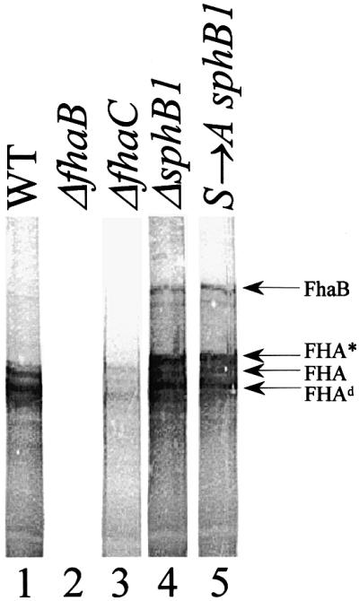
Fig. 2. Detection of cell-associated FhaB. Cellular lysates of BPSM [wild type (WT), lane 1], BPGR4 (ΔfhaB, lane 2), BPEC (ΔfhaC, lane 3), BPLC5 (ΔsphB1, lane 4) and BPLC7 (S→A sphB1, lane 5) were subjected to SDS–PAGE and immunoblotting with a mix of the anti-FHA F1, F4 and F5 antibodies. FhaB indicates the position of the FHA precursor and FHA and FHA* are as in Figure 1. Fhad represents a major degradation product of FhaB.
