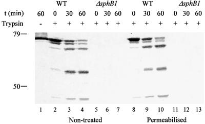
Fig. 6. Trypsin digestion of SphB1 at the cell surface. BPSM (WT, lanes 1–4 and 8–10) and BPLC5 (ΔsphB1, lanes 5–7 and 11–13) cells were treated with trypsin with (lanes 8–13) or without (lanes 2–7) a prior Tris–EDTA treatment to permeabilize the outer membrane. In lane 1, BPSM cells were mock treated for 1 h. At the indicated times, aliquots were withdrawn and trypsin was inactivated with AEBSF. The proteins were precipitated and analysed by immunoblotting with the anti-SphB1 antiserum. The masses in kDa of the molecular markers are indicated at the left.

An official website of the United States government
Here's how you know
Official websites use .gov
A
.gov website belongs to an official
government organization in the United States.
Secure .gov websites use HTTPS
A lock (
) or https:// means you've safely
connected to the .gov website. Share sensitive
information only on official, secure websites.
