
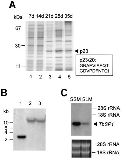
Fig. 1. TbSP1 identification. (A) Equal amounts of SLM-released protein were subjected to SDS–PAGE and stained with Coomassie Blue R-250. Days of in vitro culture (d) and the migration positions of molecular mass markers are indicated at the top and at the left, respectively. The migration position (p23) and the N-terminal sequence (p23/20) of the TbSP1 polypeptide are shown on the right. (B) Genomic DNA digested with EcoRI (lane 1), BamHI (lane 2) or HindIII (lane 3) was probed with the TbSP1 cDNA. The migration positions of DNA size markers are indicated on the left. (C) Balanced amounts of total RNA extracted from synthetic solid (SSM) or liquid (SLM) mycelial cultures were gel fractionated and probed with the 32P-labeled TbSP1 cDNA. The migration positions and the amounts of the 28S and 18S rRNAs, utilized as internal references, are shown on the right and in the lower panel, respectively.