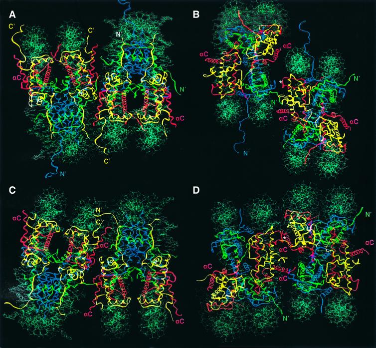
Fig. 4. Protein–protein interactions within the crystal lattice. (A) Crystal contacts between two neighboring nucleosomes in X.laevis in a view that places the superhelical axis in a horizontal orientation (as seen in Figure 1C). (B) The same Xla-NCP packing as shown in (A), but viewed down the molecular 2-fold axis. This view is achieved by a 90° rotation around the superhelical axis (horizontal). In both views, the nucleosome core particle to the left of the pair corresponds to the center green particle in Figure 3A, whereas the right-hand particle corresponds to the blue particle in Figure 3A (boxed in Figure 3A and C, respectively). Histones are colored as in Figure 1. The location of the H2A and H4 histone tails is indicated. The position of the Mn2+ ion that is crucially involved in forming crystal contacts is shown (*). (C and D) Two yeast nucleosome core particles shown in the same orientation as seen in (A) and (B), respectively. With respect to Figure 3C, the same two particles are depicted as for Xla-NCP.

An official website of the United States government
Here's how you know
Official websites use .gov
A
.gov website belongs to an official
government organization in the United States.
Secure .gov websites use HTTPS
A lock (
) or https:// means you've safely
connected to the .gov website. Share sensitive
information only on official, secure websites.
