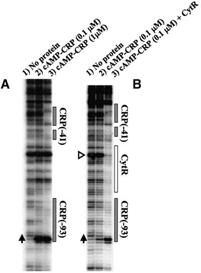
Fig. 7. cAMP–CRP binding to deoP2 DNA as probed with DNase I. The DNase I footprint was revealed by a primer extension method since a supercoiled DNA template (pdeoP2) was used (see Materials and methods). (A) Different concentrations of CRP were reacted with deoP2 in the presence of 0.1 mM cAMP. (B) cAMP (0.1 mM) and CRP (0.1 µM) were reacted with pdeoP2 in the presence or absence of CytR (15 nM). The cAMP–CRP binding site is shown as a shaded box and CytR as an open box. Vertical arrows show the hyper-reactive T at –99, and the open triangle in (B) a hyper-reactive A (–65) in the middle of the CytR binding site (see text). The deoP2 promoter sequence (–105 to –25) is 5′-AATTATTTGAACCAGATCGCATTA CAGTGATGCAACTTGTAAGTAGATTTCCTTAATTGTGATGTG TATCGAAGTGTGTTG-3′, in which the two cAMP–CRP sites are underlined.

An official website of the United States government
Here's how you know
Official websites use .gov
A
.gov website belongs to an official
government organization in the United States.
Secure .gov websites use HTTPS
A lock (
) or https:// means you've safely
connected to the .gov website. Share sensitive
information only on official, secure websites.
