
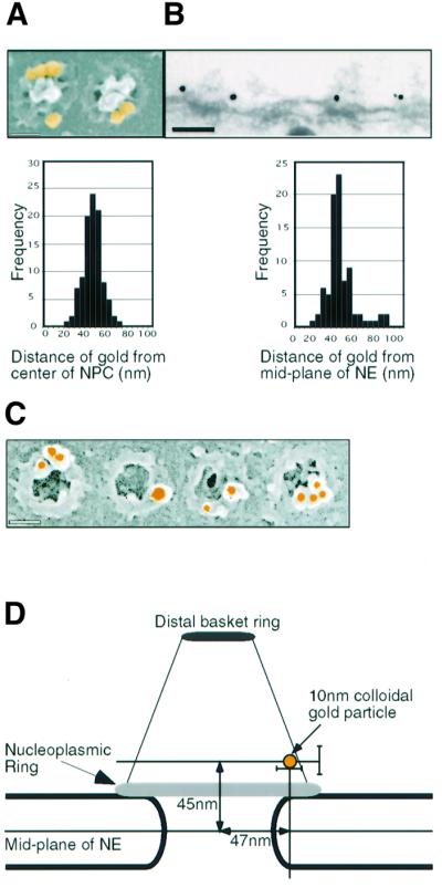
Fig. 1. Localization of Nup153 within the NPC. Fixed Xenopus oocyte nuclear envelopes were incubated with affinity-purified anti-Nup153 primary antibody and 10 nm colloidal gold labelled secondary antibodies and analysed using electron microscopy. (A) Upper panel: representative FEISEM image of NPCs decorated with gold particles (false coloured yellow). Gold particles were identified using the backscatter electron image. Lower panel: distribution of distances to the centre of the NPC (in nm) of 100 NPC-associated gold particles. Bar, 100 nm. (B) Upper panel: representative TEM image of a stretch of nuclear envelope (NE) decorated with anti-Nup153 primary antibodies and gold labelled secondary antibodies. Bar, 100 nm. Lower panel: distribution of distances to the mid-plane of the NE (in nm) of 85 NE-associated gold particles. (C) Representative images of immunogold localization of Nup153 on NPCs after stripping of the nucleoplasmic basket. Bar, 100 nm. (D) Schematic representation of the localization of Nup153-specific gold labelling on a diagram of the NPC basket. Note that two antibody molecules (not shown in the diagram) are interposed between Nup153 and the gold particle. Bars indicate standard deviations of measurement.