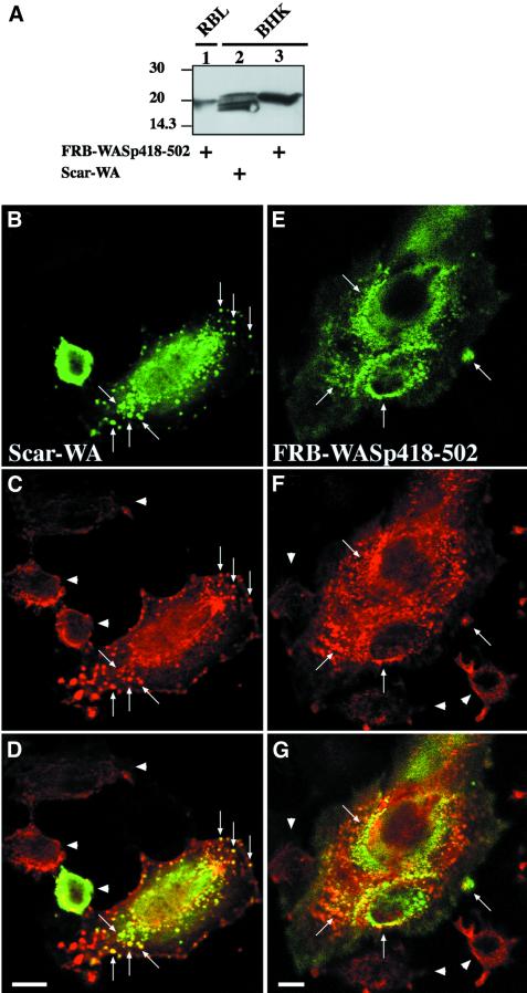
Fig. 3. Overexpressed FRB–WASp418–502 induces F-actin clusters in BHK-21 cells. (A) Lysates of RBL-2H3 cell line expressing FRB–WASp418–502, or BHK-21 cells overexpressing FRB–WASp418–502 or Scar-WA were run on 12% SDS–PAGE (9 µg total protein per lane) and blotted on to PVDF. Blots were revealed using mouse anti-myc tag antibody (clone 9E10) and the ECL procedure. The signal in RBL-2H3 cells is ∼5-fold less than in BHK-21 cells (compare lanes 1 and 3). Taking into consideration that ∼10% of BHK-21 cells expressed the FRB–WASp418–502 protein, we estimate that the level of expression of FRB–WASp418–502 is ∼50-fold lower in the RBL-2H3 stable cell line compared with that in BHK-21 cells. (B–D) BHK-21 cells expressing the Scar-WA domain. (E–G) BHK-21 cells expressing the FRB–WASp418–502 construct. (B and E) Localization of myc-tagged constructs as revealed using anti-myc tag antibody. (C and F) Distribution of F-actin. Arrows point at clusters of WASp or Scar C-terminal domains and F-actin. Arrowheads show non-transfected cells. (D and G) Superimposition of anti-myc and F-actin images. Bar: 10 µm.

An official website of the United States government
Here's how you know
Official websites use .gov
A
.gov website belongs to an official
government organization in the United States.
Secure .gov websites use HTTPS
A lock (
) or https:// means you've safely
connected to the .gov website. Share sensitive
information only on official, secure websites.
