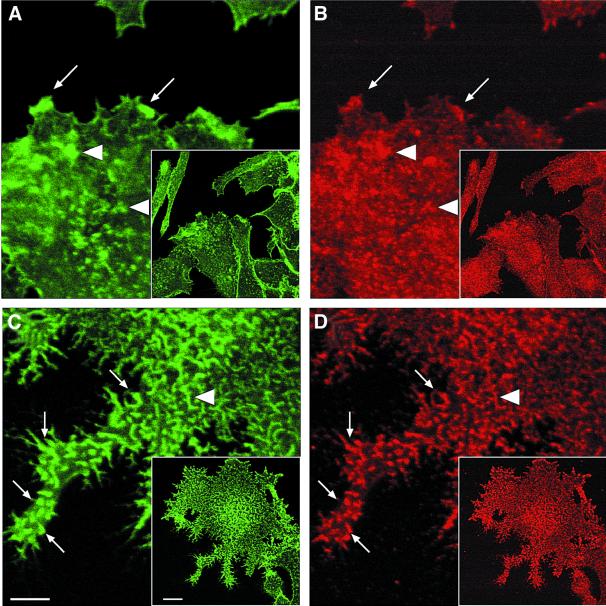
Fig. 7. Immunofluorescence localization of VASP in WASp105–502-expressing cell line. RBL-2H3 cells expressing FRB–WASp105–502 were plated on to glass coverslips coated with anti-CD25 antibodies and treated with DMSO as control (A and B) or rapamycin (C and D), fixed, and stained with phalloidin (A and C) and anti-VASP antibodies (B and D). Lower magnification fields are shown in the insets. Arrows point at peripheral structures positive for F-actin and VASP, while arrowheads depict internal structures. Confocal sectioning was performed at the ventral cell surface. Bar: 10 µm.

An official website of the United States government
Here's how you know
Official websites use .gov
A
.gov website belongs to an official
government organization in the United States.
Secure .gov websites use HTTPS
A lock (
) or https:// means you've safely
connected to the .gov website. Share sensitive
information only on official, secure websites.
