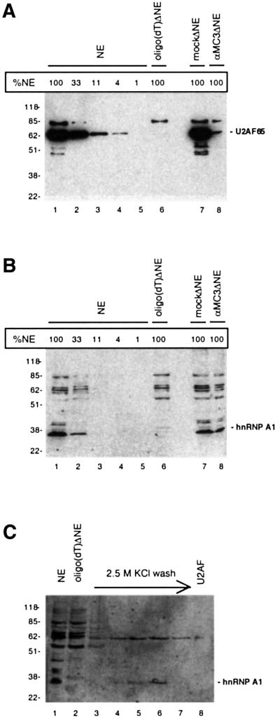
Fig. 2. Monitoring the depletion efficiency of U2AF65 (A) and hnRNP A1 [(B) and (C)] by western blotting. (A) Western blot of non-depleted extract (NE; lane 1), oligo(dT)-depleted extract [oligo(dT)ΔNE; lane 6], immunodepleted extract (αMC3ΔNE; lane 7), mock immunodepleted extract (mockΔNE; lane 8), and serial dilutions of non-depleted NE used for estimating the degree of depletion (lanes 2–5), using U2AF65-specific antibody (αMC3). Ponceau staining of the filter verified that equal amounts of extracts were loaded and transferred to the filter (data not shown). (B) Western blot of the same filter as in (A) probed with hnRNP A1-specific antibody (αA1). (C) Immunodetection of hnRNP A1 in the 2.5 M KCl buffer wash of the oligo(dT) column (lanes 3–7) and the eluted U2AF activity (lane 8). The samples were loaded next to samples containing NE (lane 1) and oligo(dT)ΔNE (lane 2). Bands above or below the marked bands probably derive from unspecific binding of the antibody. A size marker (kDa) is indicated to the left, and the positions of U2AF65 and hnRNP A1 are marked.
