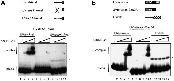
Fig. 5. Gel mobility-shift assay mapping the hnRNP A1 binding sites in the intron (A) and in the exon part of UVtat (B). The RNA substrates, indicated above the autoradiograms, were incubated in 10 µl reactions containing 0, 30, 100 and 300 ng of GST–hnRNP A1 in (A) and 0, 10, 30, 100 and 300 ng of GST–hnRNP A1 in (B). The migration of the free RNA probes and the complexes is indicated. The asterisk marks the position of RNA dimers of some of the substrates.

An official website of the United States government
Here's how you know
Official websites use .gov
A
.gov website belongs to an official
government organization in the United States.
Secure .gov websites use HTTPS
A lock (
) or https:// means you've safely
connected to the .gov website. Share sensitive
information only on official, secure websites.
