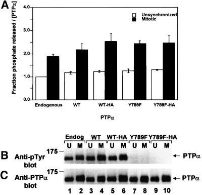
Fig. 1. Phosphatase activity of PTPα from unsynchronized and mitotic cells. Endogenous mouse PTPα (Endog) and overexpressed human PTPα (WT), PTPα-HA (WT-HA), PTPα(Y789F) (Y789F) and PTPα(Y789F)-HA (Y789F-HA) were immunoprecipitated with anti-PTPα(D2) antibody from lysates from non-overexpressor or overexpressor NIH 3T3-derived cells that were either unsynchronized (U) or arrested in mitosis (M). (The lysates were adjusted to contain approximately equal amounts of PTPα.) Aliquots of immunoprecipitates were incubated with [32P]pTyr-containing MBP in phosphatase buffer for 15 min at 30°C or subjected to anti-pTyr or-PTPα immunoblots. (A) Amount of 32P released per molecule of PTPα after 15 min incubation, normalized to the amount released by overexpressed PTPα from unsynchronized cells. Error bars indicate the SEM. (B) Anti-pTyr immunoblot of the immunoprecipitated PTPα. (C) Anti-PTPα immunoblot of the immunoprecipitated PTPα. SDS–PAGE was in 10% gels. The positions of molecular weight standards are indicated in kDa.

An official website of the United States government
Here's how you know
Official websites use .gov
A
.gov website belongs to an official
government organization in the United States.
Secure .gov websites use HTTPS
A lock (
) or https:// means you've safely
connected to the .gov website. Share sensitive
information only on official, secure websites.
