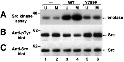
Fig. 2. Tyrosine-dephosphorylation and activation of Src in vitro by PTPα from unsynchronized and mitotic cells. Src that had been immunoprecipitated from chicken-Src overexpressing NIH 3T3-derived cells was incubated in phosphatase buffer with PTPα-HA (lanes 3 and 4) and PTPα(Y789F)-HA (lanes 5 and 6) that had been immuno purified from unsynchronized or mitotic PTPα-overexpressor cells using anti-HA antibody or with mock-immunopurified protein from control cells that did not express any HA-tagged protein (–, lanes 1 and 2). The partially dephosphorylated Src immunoprecipitates were washed to remove PTPα and then incubated with enolase and [γ-32P]ATP in kinase buffer. (A) Autoradiograph of [32P]enolase after the Src kinase assay. (B) Anti-pTyr immunoblot of the Src immuno precipitates after PTPα treatment. (C) Anti-Src immunoblot of the immunoprecipitates. SDS–PAGE was in 10% gels. The positions of molecular weight standards are indicated in kDa.
